
Behind Every Breast Cancer Diagnosis
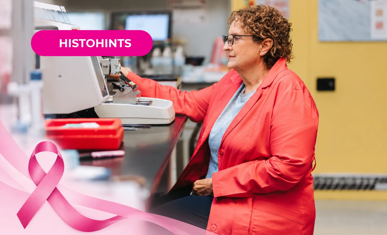
During Breast Cancer Awareness Month in October, we recognize patients and families, along with the pathologists and healthcare professionals who support breast cancer care.
But from the histology bench, awareness can look a little different.
Before a pathologist can evaluate a breast tissue specimen, there is an entire workflow behind that slide. Fixation, processing, embedding, sectioning, staining, and IHC all play a role in producing a quality slide for interpretation and diagnosis.
And as histotechs, we know that the little details can matter.
Breast cancer awareness isn’t only about the diagnosis. For histotechs, it’s also about understanding everything that happens before that diagnosis is made.
- A properly fixed specimen.
- A well-oriented block.
- A well-cut section.
- A clean, adhesive slide.
- Consistent staining.
- A coverslip without bubbles or debris.
Every step matters. And when something goes wrong in the workflow, it can create additional work, compromise slide quality, or potentially contribute to delays in a diagnosis.
A few things to watch for:
- Inadequate fixation: Follow your lab’s SOPs and CAP/CLIA guidelines for proper tissue fixation. Adequate fixation is the first step in preserving tissue morphology and supporting downstream testing.
- Processing issues: Ensure all tissues are placed on the appropriate processing schedule and that your processors are properly maintained and monitored.
- Poor embedding orientation: Proper orientation helps to ensure adequate sectioning. Follow tissue-specific needs and general orientation guidelines when embedding.
- Microtomy issues: Thick or thin sections, wrinkles, folds, chatter, or tissue wash can all affect section quality and may make interpretation more difficult.
- Staining or background issues: Inconsistent staining, excessive background, debris or other staining artifacts can interfere with the quality and clarity of the final slide, especially in digital pathology.
- Slide labeling and identification: Accurate, legible, and durable specimen identification is essential throughout the workflow. Whether it’s a cassette or stained slide, identification errors can create patient safety concerns and potentially impact a diagnosis if the specimen cannot be confidently connected to the right patient.
So where do the products we use fit into the workflow?
The right tools can’t replace good technique or a well-controlled process, but they can help support consistency at every step—from sectioning to slide preparation to staining and coverslipping.
Sectioning: A quality blade, such as PolyCut Plus, can help support consistent sectioning and efficient microtomy.
Slides and adhesion: Quality slides and coverglass, like KT Slides + Koverglass, can support tissue adhesion, slide clarity, and the digital pathology workflow.
Specimen identification: Durable, legible specimen identification is essential throughout the workflow. Cassette and slide printers, such as PiSmart, can help provide reliable identification from the cassette to the final slide.
Recent Posts
-

Behind Every Breast Cancer Diagnosis
During Breast Cancer Awareness Month in October, we recognize patients and families, along with the
-

HT or HTL? Which path is right for you?
You like histology. You’ve been working in the lab, and now you’re starting to think about getting c
-
StatClick Prefill Handling and Closure
How to properly handle and close the StatClick Prefill for leak-proof transport. Follow your facili