
Amyloid Sections Washing Off? Let’s get to the bottom of this tricky stain.

Have you ever wondered how an 8–10 µm amyloid section washes off with aggressive alkaline rinses of a Congo Red stain? If you've been in histology long enough, you know this frustration. The good news: Its most likely the slide and the drying technique.
The Amyloid Challenge: Because amyloid sections are cut twice as thick as standard H&Es, they are heavy and stiff. Without that ionic "Velcro," the edges curl, allowing reagents to seep underneath and lift the whole section.
The Slide Fix: Standard glass slides are NOT enough. You need to use an adhesion Slide (also referred to as charged, positive, plus, coated – they all mean the same thing), preferably the KT™ Slides. These slides provide a permanent positive charge that “anchors” the tissue to the slide. Without a positive slide, the tissue will wash off due to the thickness of the section.
“The Steam Engine" effect is the #1 reason for tissue loss in amyloid staining. Because 8-10 µm sections are like thick blankets, they hold much more water than a standard 3 µm H&E.
The Problem: Thick amyloid sections trap water pockets. When you hit the drying oven, that water turns to steam, creating a "blister" that pops the tissue off the glass.
Resolution: It is so important to make sure to always drain the slides vertically for at least 10 minutes before baking. This lets gravity pull the water out so the tissue can physically touch the charged glass. If you skip this, you’re essentially "boiling" the tissue off the slide.
The "Expiration" Myth
The charge on "Positive" slides can degrade over time, especially in high humidity. If your thick sections are suddenly falling off and you’re using an old box of slides, the glass—not your microtomy—is likely the culprit.
The “Apple Green” Myth
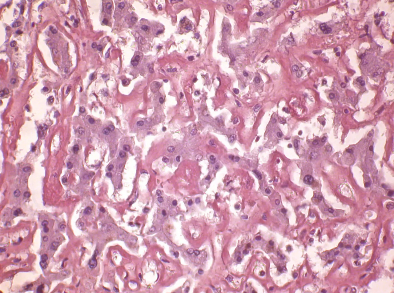
Don’t Chase the green – Chase the birefringence! Here’s the deal; a lot of people were taught that amyloid must show the classic apple green birefringence. But that’s not actually true. Section thickness, polarization angle, and stain quality can make the colors look different. Focusing only on that “Perfect” green apple color is an easy mistake. You might see green, but also yellow, blue, orange, or even a mix. Put simply, if it is birefringent, it can still be amyloid.
Related Products
Recent Posts
-

Amyloid Sections Washing Off? Let’s get to the bottom of this tricky stain.
Have you ever wondered how an 8–10 µm amyloid section washes off with aggressive alkaline rinses of
-

Thinking of going xylene-free? Here’s what labs should know.
Histotechs know the dangers and health risks associated with xylene, and interest in greener labs co
-

Manual vs. Semi-Auto Microtomes: Why the small difference makes a big impact.
Manual microtomes have long been a reliable choice in histology labs, but semi-automatic models, lik
