
KT Slides & Background Staining

Background
Whole slide imaging (WSI) is gaining widespread adoption in diagnostic and research pathology. While attention is often given to image clarity and scanner performance, the microscope slide itself can be an influential factor in digital pathology.
Background staining, an artifact arising from residual stain retained on slide surfaces, can affect the efficiency and economics of digital scanning systems as it can introduce a non-tissue signal that scanners interpret as tissue. This study evaluates the impact of background staining to whole slide image file size and scanning speed between 2 different brands of adhesion slides.
Results
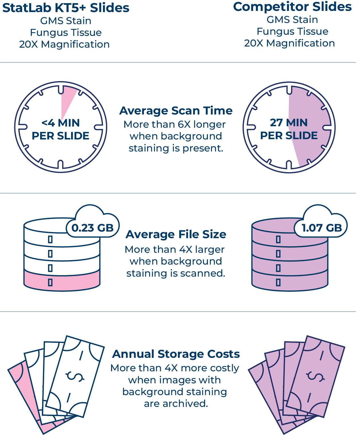
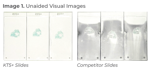
In this initial focused study, the evaluated competitor adhesion slides exhibited moderate to severe background staining, resulting in significantly longer scan times and file sizes, whereas the StatLab KT5+ slides exhibited minimal background staining, resulting in quicker scanning times and smaller file sizes.
Materials
StatLab KT5+® Premium Advanced Adhesion Slides
Competitor Adhesion Slides
StatLab MasterTech™ GMS (Grocott’s Methanimine Silver) Stain Kit
Motic®EasyScan One Digital Imager, EasyScan Application version 1.0.7.156
StatLab Acrymount™ Plus Mounting Medium
StatLab KT® Koverglass Thickness #1, 24 x 50 mm
Methods
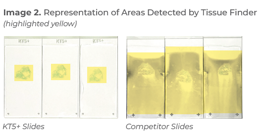
Twenty-five slides of each slide brand were prepared using 4 μm sections of formalin-fixed, paraffin-embedded tissue containing known positive fungal elements from the same tissue block, manually stained with the same GMS special stain protocol, coverslipped, and dried overnight at ambient temperature. Each slide was then scanned at 20x magnification using a high-resolution whole slide imager according to the manufacturer’s manual. The tissue finder was set to auto-scan per the manufacturer’s manual. Metrics evaluated included total scan time per slide and whole slide image file size. This study did not evaluate
clinical diagnostic quality or outcomes.
Conclusion
The results demonstrate that slide choice not only directly affects background staining, which is not simply a visual concern, but also contributes to workflow inefficiencies. For labs scaling digital pathology programs, these inefficiencies could affect turnaround times, storage infrastructure, and long-term costs.
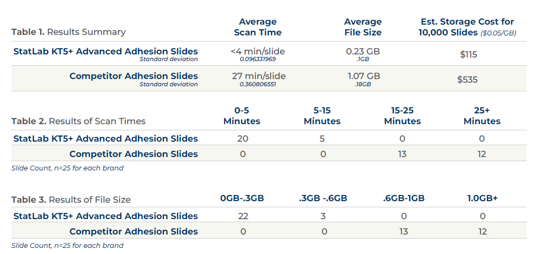
Slides with moderate to severe background staining required six times longer to scan (Table 1). Scan times could be reduced by manually adjusting the tissue finder for each slide or utilizing ‘Preview Mode’ but that would increase the hands-on time of histotechs, potentially informing the headcount needs of a lab.
File sizes of scans, including background staining, were more than four times as large as slides that had minimal background staining. In the December 2025 issue of Laboratory Economics1, Dr. Lee Cooper, Director of the Institute for Artificial Intelligence in Medicine and a professor of pathology and toxicology at Northwestern University Feinberg School of Medicine, stated file size for a “typical slide is about 1 GB, and that cloud storage costs could be between $.01 and $.07 per GB per year.” A large lab that is looking to adopt a full digital workflow can readily store between 1,000 GB and 1,000,000 GB.2
Using estimated costs of $.05 GB annual storage rate per slide, a lab’s storage cost for 10,000 slides without background staining, like the StatLab KT5+, would be $115, whereas a slide with moderate to severe background staining averaging 1GB in size would be ~ $535 for the same number of slides, which is an increase of nearly four times in cost comparison.
Choosing a digital-ready slide, such as the StatLab KT5+, that exhibits minimal background staining is a practical and actionable way for labs to improve consistency, efficiency, readiness for increasing digital workflows, and remove barriers to digital adoption.
References
- Cooper L. [What is the Future of Artificial Intelligence in Digital Pathology?]. Laboratory Economics. December 2025.
- Lars Ole Schwen, Tim-Rasmus Kiehl, Rita Carvalho, Norman Zerbe, André Homeyer, Digitization of Pathology Labs: A Review of Lessons Learned, Laboratory Investigation, Volume 103, Issue 11, 2023, 100244, ISSN 0023-6837, https://doi.org/10.1016/j.labinv.2023.100244.
The findings and conclusions presented are based on internal research conducted by StatLab Medical Products under specific testing conditions. These results should be considered preliminary and are not intended to replace independent external studies or peer-reviewed research. Performance may vary depending on laboratory protocols, equipment, environmental factors, and specimen characteristics.
StatLab Medical Products makes no warranties, express or implied, regarding the completeness, accuracy, or reliability of the information provided. The mention or display of third-party products is for comparative purposes only and does not imply endorsement or criticism. Readers are encouraged to conduct their own evaluations and consult appropriate regulatory and scientific sources before making purchasing or clinical decisions.
StatLab Medical Products shall not be held liable for any direct, indirect, incidental, or consequential damages arising from the use or reliance on the information contained in this document.
Recent Posts
-

KT Slides & Background Staining
Background Whole slide imaging (WSI) is gaining widespread adoption in diagnostic and research path
-

Happy National Histotechnology Professionals Day!
Thank you for the vital role you play in providing the best possible patient care. We see your hard
-

Why HCA Mid-America Division only uses KT slides in their lab.
One lab's story of overcoming tissue wash challenges.
